Surgical navigation systems optics and cancer

According to the functions of the optical surgical navigation, it allows the detection of cancer in patients, also decreases the arrival times to the specialist of the patient, by means of guidance to identify and overcome the barriers that face and prevent early detection, as well as adequate and timely diagnosis and treatment of the disease. In addition, health experts achieve levels of accuracy and security in image acquisition, which are extended and assisted by computer, magnifying images, facilitating diagnosis, planning and execution of procedures.
What are the benefits of optical surgical navigation system?
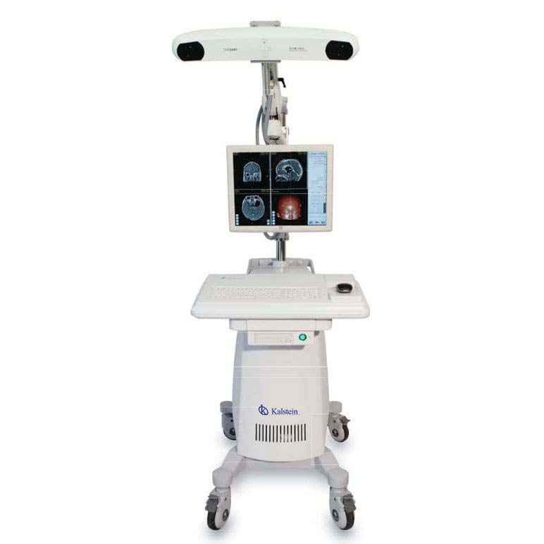
Through the use of optical surgical navigation systems, it allows the development of surgeries by means of real augmented technological simulation, which is characterized by sensory perception, adding visual content, as help guides to make them less invasive and show the role of the staff as comparative advantage over virtual reality.
Laminar Flow Campaigns and Biological Safety?
In professional practice and for many laboratory jobs, it is important to protect the product from pollution, whether from the environment or from personnel. When working with hazardous substances, it is necessary to protect the life of the operator, and to achieve this, laminar flow hoods are ideal, these allow to protect the product with which you are working. As well as biological safety, it preserves the product, the personnel and the environment.
What are the Applications for High Speed Centrifuge and Low Speed Centrifuges?

Generally, centrifuges are equipment that form rotating movements, managing to separate elements in the samples for study separately, either by decanting or sedimentation. These equipment are used in laboratories, becoming fundamental devices, adapted to the different procedures in the working environment.
Care and maintenance of an ultrasound scanner?
Ultrasound scanners must be maintained to be handled correctly, so care must be reasonable by inspection, sterilization or disinfection, as necessary. Failure to comply with the care and maintenance of the equipment can cause difficulties in the analysis of the same, showing undesired and erroneous results, causing irreparable damage of the equipment.
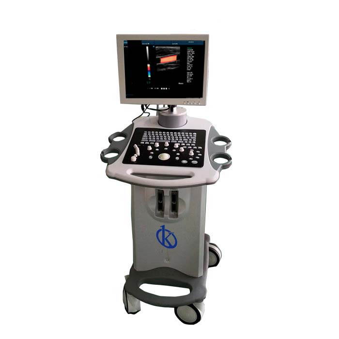
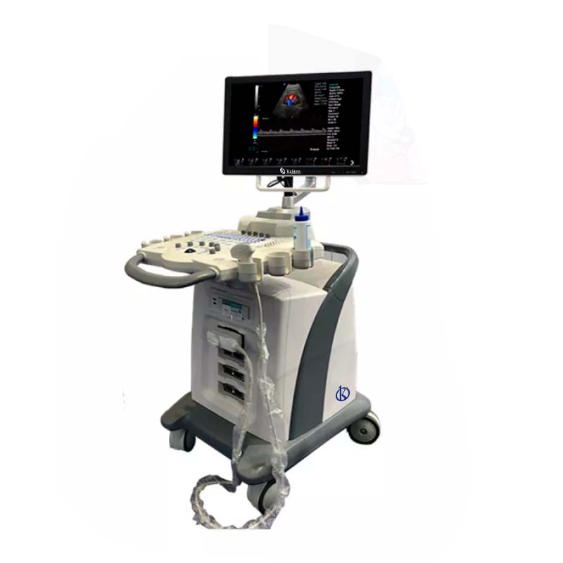
What is an ultrasound scanner?
The ultrasound scanner is a medical equipment of general use, in which it allows the obtaining of abdominal, gynecological-obstetric images, small parts, vascular, as well as for cardiology and portable type, and intravascular images. Some means include attached transducers to provide more specialized programming, such as heart, vascular, endovaginal, endorrectal, or small-part scanning (e.g., thyroid, breast, scrotum, prostate).
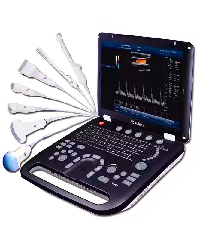
For use with a portable ultrasound scanner?
This handheld ultrasound device is small and very low cost, has as advantages that makes ultrasound more simple, also allows you to be everywhere inside the hospital, facilitating the doctor’s tour of the patient’s body; it is used in a variety of applications in different departments, the widest for a single ultrasound probe, making it an imaging device for the whole body, in urological, abdominal, fetal, cardiovascular, gynecological and musculoskeletal applications, among others.
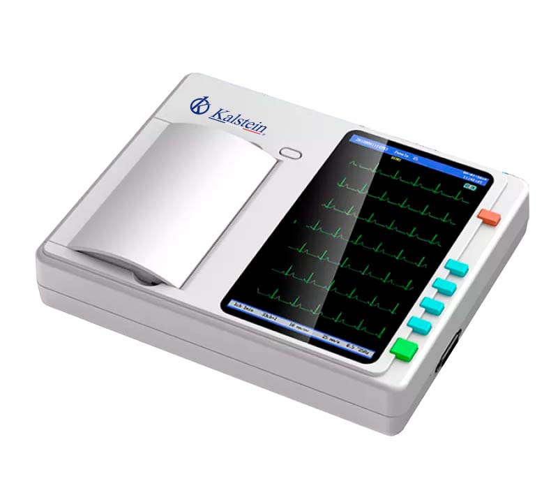
Care and maintenance of an electrocardiograph

An electrocardiograph is a medical device that analyzes the electrical mechanisms of the heart over a period of time. It works in conjunction with electrodes placed on the patient’s chest and limbs to detect electrical changes.
What are the differences between a mono channel and multichannel electrocardiograph?
The electrocardiograph is a medical team in charge of performing tests called electrocardiogram (ECD), and detect the signals of the heart, and in turn, to diagnose some of the diseases of the heart. This device is essential in all health centers, especially in cardiology units.
What does an electrocardiograph represent?

This device that captures and expands the electrical activity of the heart through electrodes, this device represents the electrical activity of myocardial fibers, produced when cardiac muscles change their electrical properties with each beat, then electrodes attached to the body can be used, to measure changes in the body’s voltage, and this is called electrocardiogram.